

Translate this page into:
Three-dimensional canine loop for management of buccally erupted canines
This article was originally published by Wolters Kluwer and was migrated to Scientific Scholar after the change of Publisher.
Abstract
Maxillary canines are known as the cornerstones of mouth. They are considered to be important for esthetics and for functional occlusion. Any disturbance in the eruption process leading to an aberrant position will hamper esthetics as well as function. Orthodontic tooth movement of total buccally blocked-out canine is usually difficult as it is related with the problems of severe crowding, midline deviation, involvement of long root movement and risk of gingival recession. Such conditions can be treated orthodontically in various ways, but this clinical innovation helps to correct the buccally placed canines into the arch with a precise control of the canine in all the Three-dimensions (3D) of space as well as providing maximum comfort to the patient by placing the canine loop on the palatal surface of the tooth, reducing soreness on the labial mucosa. It can be easily fabricated and activated at chairside for either simultaneous or sequential control in 3D.
Keywords
Buccally placed canines
loops
ectopic canine

INTRODUCTION
The prevalence of permanent maxillary canine impaction or ectopic eruption in the general population is approximately 1-2%.[1,2] Palatally displaced canines occur twice as frequently as buccally.[3] However, buccally displaced canines are commonly seen in practice. Placement of an attachment over buccally placed canines [Figure 1] usually causes ulcerations on cheeks to patients who are susceptible to recurrent episodes of mouth sores, which may be precipitated or exacerbated by irritation from them. This is not an emergency but may be very uncomfortable for the patient. Placing wax over the cuspid bracket in the area of irritation may also bring relief, and allow the irritated area to recover.

- Highly placed canine
The current scenario in orthodontics demands minimal discomfort to patients, and hence an innovative clinical technique was devised to treat buccally erupted canines high in the vestibule by placing the bracket palatally [Figure 2].
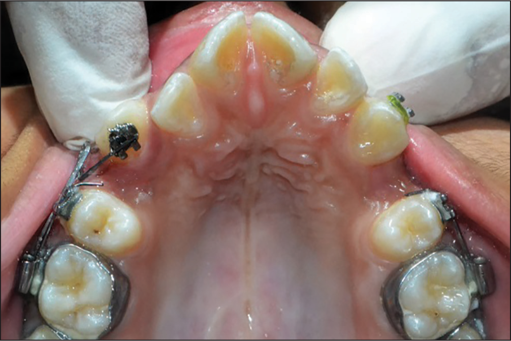
- Placement of orthodontic bracket on the palatal aspect in buccally placed canine
Steps for fabrication of three-dimensional canine loop [Figure 3]:

- Steps in fabrication of three-dimensional canine loop
Placement of an edgewise bracket on the palatal side of the buccally placed canine (to minimize the soreness and ulceration caused by buccal placement of the bracket thereby decreasing the discomfort to the patient).
A closing loop is bent on a sectional 0.017″ × 0.025″ TMA with mesial vertical arm of 6 mm and distal vertical arm of 13 mm with the distance between the legs of the loop kept at 2 mm. The distal vertical arm is kept longer than the mesial vertical arm to aid in extrusion of the highly placed canine into the arch. The height of the distal vertical arm can be adjusted depending on the amount of extrusion required. Distal vertical arm is bent toward the tissue surface for ease in placement of the three-dimensional (3D)-Loop over the palatally placed cuspid bracket.
A 90° bend is given to the distal vertical arm to form the active arm, which will engage the slot of the palatally placed cuspid bracket in an edgewise mode.
Pre activation palatal root torque of 15° is given to the active arm to maintain the roots of cuspids in the cancellous bone thereby avoiding cortical anchorage and the possibility of dehiscence during movement of the labially placed cuspid.
A mesial up or a mesial down bend of 15° can be given on the active arm to allow angulation correction of the labially placed cuspids.
The mesial vertical arm is bent 90° distally to insert into the auxiliary slot of the molar tube.
A 3D Canine loop was placed in a patient with ectopically erupted canine and results were found to be favorable [Figures 4-9].
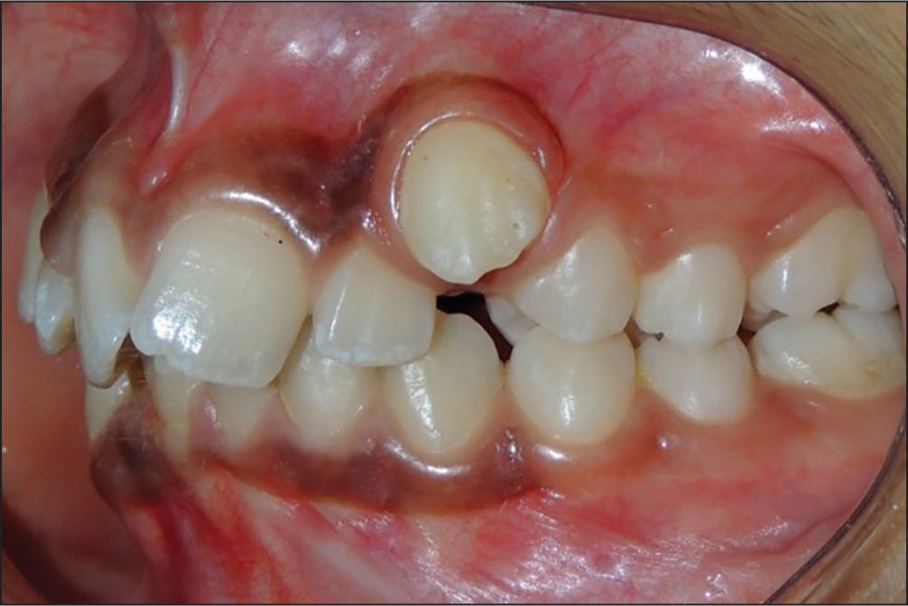 Figure 4
- Buccally placed canine
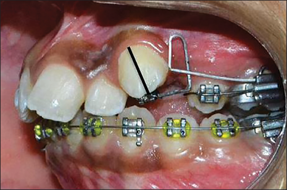 Figure 5
- Placement of three-dimensional canine loop on the palatal aspect
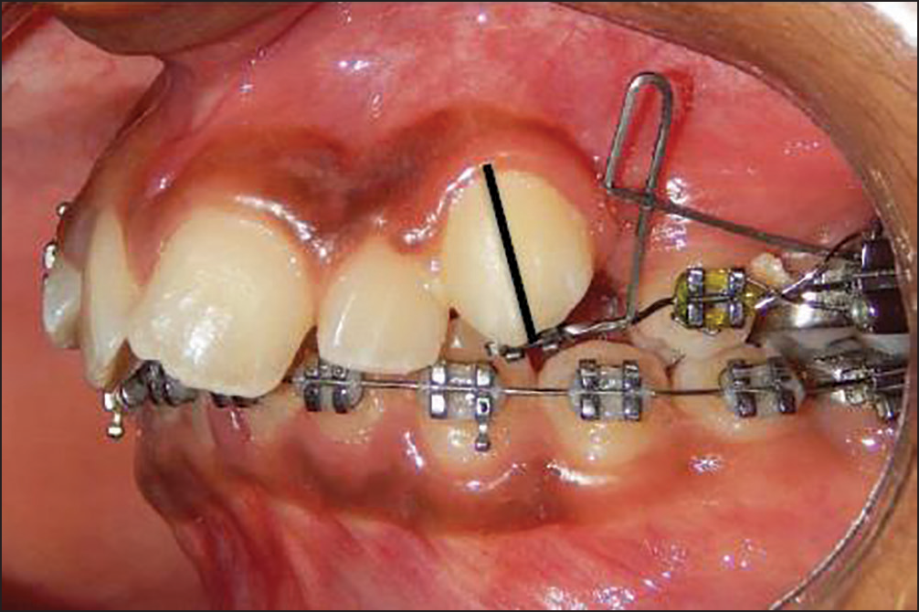 Figure 6
- Descent of buccally placed canine; correction in angulation with maintenance of torque
 Figure 7
- Buccally placed canine in the line of occlusion with uprighting of canine and maintenance of torque
 Figure 8
- Continuous arch mechanics
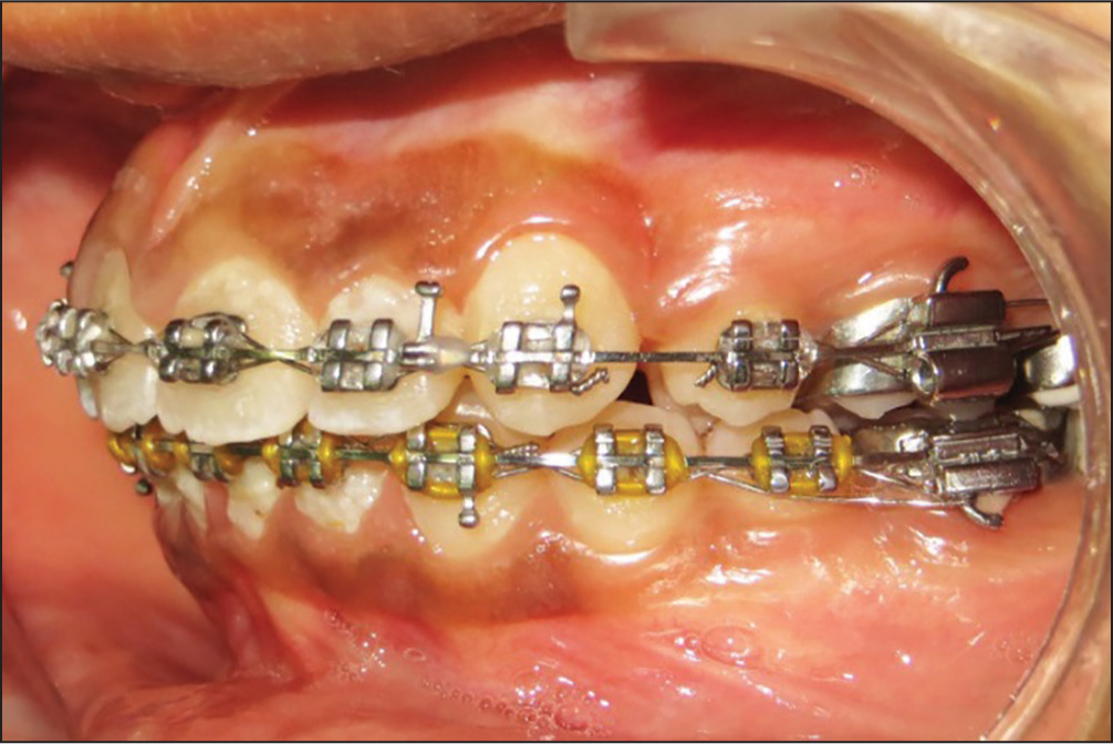 Figure 9
- Correction of Buccally placed canine
CONCLUSION
Movement in all the three planes of space is possible with 3D canine loop (tip, torque and extrusion) along with less discomfort to the patient since the attachment is placed on the palatal aspect of labially placed canine thereby reducing ulceration on the mucosa of the lips and cheeks.
Financial support and sponsorship
Nil.
Conflicts of interest
There are no conflicts of interest.
References
- A review of the diagnosis and management of impacted maxillary canines. J Am Dent Assoc. 2009;140:1485-93.
- [Google Scholar]
- Influence of radiographic position of ectopic canines on the duration of orthodontic treatment. Angle Orthod. 2009;79:442-6.
- [Google Scholar]
- Canine impactions: Incidence and management. Int J Periodontics Restorative Dent. 2006;26:483-91.
- [Google Scholar]


