

Translate this page into:
Evaluation of smile esthetics in Central India
This article was originally published by Wolters Kluwer and was migrated to Scientific Scholar after the change of Publisher.
Abstract
Objectives
This study was undertaken: (1) To establish static norms in the central India population for the various smile parameters. (2) To analyze and quantify the sexual dimorphism of esthetic smile. Settings and Design: Hundred subjects (50 males and 50 females) with an average age of 14.5 years with a pleasing smile were selected for the study. Static photographs with posed smile in natural head position (NHP) were taken.
Materials and Methods
Following smile parameters were quantified using Adobe Photoshop ruler software: Maxillary incisor exposure (MIE) (mm), smile index (SI) (mm), smile arc, buccal corridor ratio (%), Most posterior maxillary tooth visible, anterior height of the smile (%), posterior height of the smile (%).
Statistical Analysis
The data were compiled systematically. Statistical analysis was done using Statistical Package of Social Sciences (SPSS version 17.0). For MIE and SI, Unpaired t-test was applied. Chi-square test was applied for most posterior maxillary tooth visible, smile arc buccal corridor, anterior height of smile, and posterior height of smile.
Results
The mean value of MIE was found to be 7.76 mm and 8.82 mm in boys and girls, respectively. SI showed a mean value of 8.26 for boys and 7.91 for girls. Girls displayed second premolar commonly in contrast to boys, who displayed first premolar. Parallel smile arc was noted more frequently in females, while males displayed flat smile arc commonly.
Conclusion
Orthodontists, being smile architects, have the responsibility to design and create smiles. Smile analysis should be an integral part of dental/orthodontic treatment planning and mechanotherapy.
Keywords
Smile analysis
smile index
esthetic smile

INTRODUCTION
The specialties of orthodontics have experienced the re-emergence of the soft tissue paradigm in recent years with greater focus on soft tissues around the mouth in general and smile in particular in diagnosis and treatment planning.[1] Smile is defined as facial expression characterized by upward curving of the corners of the mouth.[2] Smiles are classified as stages I and II.[2] Stage I smile is a posed smile, while stage II is an unposed (spontaneous) smile.[3]
The posed social smile is repeatable photographically in comparison with Duchenne (enjoyment) smile.[4] Most studies refer to the posed smile as it is reproducible and can be used as a reference position.[5]
The subject of smile and facial animation is of great interest to orthodontists. Since the mouth and teeth play a key role in facial esthetics,[2] orthodontists have to make every effort to develop a harmonious balance between the various soft and hard tissue structures that will produce an attractive smile.[6] This will be possible only when they are aware of the principles that manage the balance between the teeth and soft tissues during a person’s smiles.[7,8] Lack of this knowledge in treatment planning and mechanics can cause a flattening of the smile arc during orthodontic treatment,[3] thus creating less esthetic smiles. Therefore, it is recommended that a frontal smile photograph be used as a standard orthodontic clinical record to aid in initial diagnosis and treatment planning.[9-12]
During clinical examination emphasis is placed on the display zone of smile, which is determined by lip thickness, intercommissural width, interlabial gap, smile index (SI), and gingival architecture.[4] Inclusion of smiling photographs, with the usual frontal and lateral photographs makes it possible to observe patients in a much more natural attitude. Although various scientific studies examined smile esthetics using static photographs to determine relationships and proportions,[1,2,4] few studies have been reported in the Indian population.
This study was undertaken with the following aims and objectives:
-
To establish static norms in the Central India population for the following smile parameters:
Maxillary incisor exposure (MIE) (mm)
Smile index (mm)
Most posterior maxillary tooth visible
Smile arc e. Buccal corridor ratio (%)
Anterior height of the smile (%)
Posterior height of the smile (%).
To analyze and quantify the sexual dimorphism in the various components of esthetic smile.
MATERIALS AND METHODS
Hundred subjects (50 boys and 50 girls) [Table 1] aged about 13-16 years [Table 2] with a pleasing smile from various schools in Bhopal were selected for the study. A stratified random sampling design was used to collect sample. Out of several schools present in each zone, one has been selected on a random basis through lottery method. This ensures that each child between thee age group 13 and 16 years has got an equal chance of getting selected in the sample. The sample size has been taken on the basis of the pilot study.
| Sex | No. of patients | Percentage |
|---|---|---|
| Boys | 50 | 50 |
| Girls | 50 | 50 |
| Total | 100 | 100 |
| Age group (years) | No. of subjects |
|---|---|
| 13-14 | 15 |
| 14-15 | 55 |
| 15-16 | 30 |
| Total | 100 |
Written consent from the schools and the parents were taken before proceeding with the study. Ethical clearance for the study was taken from Ethical Committee of People’s College of Dental Sciences and Research Center. The selected samples had Angle’s class I molar relationship with complete permanent dentition up to the second molars present. They had no previous history of orthodontic treatment, maxillofacial surgery, restoration/ prosthetic crowns in the anterior teeth, or periodontal treatment (except for routine scaling and polishing). Clinical examination was carried out, and upper and lower impressions were made to prepare study models for records.
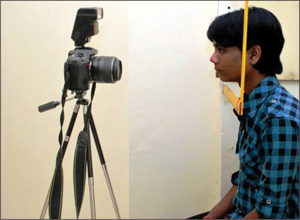
Static photographs with posed smile in natural head position (NHP) were taken. All photographs were taken in a similar environment and lighting conditions using Nikon D-60 SLR camera, which was mounted on a tripod stand at a fixed distance of 20 inches [Figure 1]. Focal length of 38 mm was set. The lens was positioned parallel to the true perpendicular of the face in NHP, and the camera was raised to the level of patient’s lower facial third. The patient was asked to say “cheese” and then smile. The photographs were then transferred to computer software (Adobe Photoshop 7.0.1) and were cropped with vertical (tip of the nose and soft-tissue pogonion and perpendicular drawn from the zygomatic prominence) limits. All images were then adjusted to a standardized image size. Measurement between two points (subnasale to soft tissue menton) was considered representative to check magnification error. This was then compared with clinical measurements and was found to have a statistically significant correlation. The Adobe Photoshop ruler software was used to obtain measurements for this study. The following data were recorded:
- Smile capture method
Maxillary incisor exposure [Figure 2] — amount of vertical display of the maxillary central incisors was measured in mm. The Adobe Photoshop ruler software was used to measure MIE
 Figure 2
- Maxillary incisor exposure
-
Smile index [Figure 3] — was described by Ackerman et al. (1998)[3] as:
Intercommissural width on smiling — with the ruler tool in Adobe Photoshop a horizontal line was drawn from the corner of the lips on one side to the same point on the contralateral side. The distance between the two points was measured.
 Figure 3
- Smile index
Interlabial gap on smiling — the distance in mm between the upper and lower lips at midline. The Adobe Photoshop ruler software was used to measure distance between the upper and lower lips.
Most posterior maxillary tooth visible — entered either as canine, first premolar, second premolar, or first molar. In the case of a discrepancy between the two sides, the most posterior tooth was entered
Smile arc [Figure 4] — was entered as parallel (when the incisal edges of the maxillary anterior teeth followed the curvature of the lower lip), flat (when the incisal edges of the maxillary anterior teeth had no curvature relative the lower lip line), or reverse (when the incisal edges of the maxillary anterior teeth had a reverse curve relative the lower lip line)
 Figure 4
- Smile arc
Buccal corridor ratio [Figure 5] — for this measurement, a horizontal line was drawn from the most posterior maxillary tooth on one side to the same point on the contralateral side (maxillary interdental width). A second line was drawn from the narrowest point visible in the inner commissure of the buccal mucosa to the same point on the opposite side. The buccal corridor ratio was calculated according to the method given by Frush and Fisher (1958)[13] as:
 Figure 5
- Buccal corridor ratio
Anterior height of the smile [Figure 6] — entered as either high smile (a contiguous band of gingiva above the maxillary central incisor), average smile (showing 75% to 100% of the maxillary central incisors), or low smile (showing <75% of the maxillary central incisors)
 Figure 6
- Anterior height of the smile
Posterior height of the smile [Figure 7] — entered as either high smile (a contiguous band of gingiva above the maxillary first premolar), average smile (showing 75% to 100% of the maxillary first premolar), or low smile (showing <75% of the maxillary first premolar visible).
 Figure 7
- The posterior height of the smile
RESULTS
Data were statistically analyzed using Statistical Package of Social Sciences (SPSS version 17.0 manufactured date 6-5-2010). For MIE and SI, unpaired t-test was applied. Chi-square test was applied for most posterior maxillary tooth visible, smile arc, buccal corridor, anterior height of smile, and posterior height of smile [Table 3].
| Component | Gender | No. of Samples (n) | P value | Result |
|---|---|---|---|---|
| MIE | Boys | 50 | 0.1620 | NS |
| Girls | 50 | |||
| SI | Boys | 50 | 0.3292 | NS |
| Girls | 50 | |||
| MPMTV | Boys | 50 | 0.0150 | S |
| Girls | 50 | |||
| Smile Arc | Boys | 50 | 0.27376095 | NS |
| Girls | 50 | |||
| Buccal Corridor Ratio | Boys | 50 | 0.06551871 | NS |
| Girls | 50 | |||
| AHS | Boys | 50 | 0.00460092 | S |
| Girls | 50 | |||
| PHS | Boys | 50 | 0.01916825 | S |
| Girls | 50 |
Maxillary incisor exposure
The mean value of MIE was 7.76 mm in boys and 8.18 mm for girls. Unpaired t-test was applied, and P value was calculated. The P value showed that there is no significant difference between boys and girls [Table 3].
Smile index
The mean value of SI was 8.26 mm in boys and 7.90 mm in girls. Unpaired t-test was applied, and P value was calculated. The P value showed that there is no significant difference for SI between boys and girls [Table 3].
Most posterior maxillary tooth visible
Fifty-eight percent of the samples displayed the second premolar most posterior maxillary tooth visible on smile, whereas 42% displayed the first premolar [Figure 8]. There was a significant difference between boys and girls. Girls (70%) showed second premolar tooth more commonly, whereas first premolar tooth was more commonly displayed by boys (54%) [Figure 8]. None of the subjects showed canine or first molar as most posterior maxillary tooth visible. Chi-square test was applied, and P value came out to be significant. P value showed that there is a significant relationship between gender and most posterior maxillary tooth visible.

- Gender differences in most posterior tooth visible
Smile arc
Forty-six percent of the sample showed flat smile arc, 45% a parallel smile arc, and 9% a reverse smile arc [Table 4]. Flat smile arc was commonly seen in boys (54%), whereas girls (52%) showed parallel smile arc [Figure 9]. Chi-square test was applied, and P value was found to be nonsignificant. The P value showed that there is no significant relationship between gender and smile arc.

- Gender differences in smile arc
| Smile Arc | Boys | Girls | Total |
|---|---|---|---|
| Parallel | 19 | 26 | 45 |
| Flat | 27 | 19 | 46 |
| Reserve | 04 | 05 | 09 |
| Total | 50 | 50 | 100 |
Buccal corridor ratio
Most frequent buccal corridor was that of medium type and was found in 50% cases, second frequent category was of medium-broad type constituting 26% cases. Twelve percent of total showed broad smile fullness and 10% showed medium-narrow smile fullness. Narrow smile fullness was seen least commonly in two percent of cases [Figure 10]. Medium smile fullness was most commonly found both in boys and girls, seen in 62% of boys and 38% girls. Medium-broad smile fullness was seen in 30% girls and 22% boys. Broad type was seen more commonly in girls (20%) than in boys (4%). Medium-narrow and narrow type were equally found in girls and boys constituting 10% and 2% each, respectively [Figure 10]. P value showed that there is no significant relationship between gender and buccal corridor [Table 3].

- Gender differences in buccal corridor ratio
Anterior height of the smile
Sixty-four percent subjects showed average anterior height, followed by 30% subject showing a low anterior smile height. High anterior smile height was seen in six subjects. Seventy-four percent girls showed an average anterior smile height. Fifty-four percent boys showed average anterior height smile, while 44% showed a low anterior smile height [Figure 11]. Chi-square test was applied, and P value was found to be significant. The P value showed that there is a significant relationship between gender and the anterior height of the smile [Table 3].

- Gender differences in anterior height of the smile
Posterior height of the smile
Forty-seven percent showed average posterior height of smile, 44% showed low type and 9% showed high posterior height of smile. In boys, low posterior height of smile was more commonly seen (54%), followed by average posterior height of smile in 44%. Girls showed average posterior height of smile more commonly (50%) than low posterior height of smile (34%) [Figure 12]. Chi-square test was applied, and P value was found to be significant. The P value showed that there is a significant relationship between gender and the posterior height of the smile [Table 3].

- Gender differences in posterior height of the smile
DISCUSSION
The present study was undertaken to evaluate various parameters in esthetic smile. In the present study, static photographs of posed smile were taken in frontal view with NHP,[4,14-16] whereas couple of studies recorded and evaluated dynamic smile in addition to static evaluation.[17,18]
The most frequent type of smile is the average smile that reveals 75-100% of maxillary incisor length. Geron and Atalia[19] have concluded that 1 mm of upper-gingival exposure at smile and speech is within the esthetic range. However, smiles with excessive upper and lower gingival display are considered less attractive.[19] In the present study, mean value of MIE at smile was 7.76 mm for males and 8.18 mm for females. Sixty-four percent of the subjects had average anterior smile height, out of which 27% were boys, and 37% were girls, which is comparable to other studies.[16] Our results agree with that of Tjan et al.[20] who found a sexual dimorphism in that low-smile lines are a predominantly a male characteristic (2.5:1; M:F) and high-smile line is predominantly a female characteristic (2:1; F:M). The present study showed that the low-smile line is common in males (M:F = 2.7:1), while average smile line was more commonly observed in females.
The mean value for SI was 8.26 mm for boys and 7.91 mm for girls. No statistical significance was found between gender and SI. Ackerman et al.[21] reported a SI of 6.04 mm for boys and 6.29 mm for females. They also found no statistical significance between gender and SI. Schabel et al.[22] noted a mean value of 5.3 mm for SI in their study.
A key component of an esthetic smile is a consonance between the arcs formed between the incisal edges of the maxillary teeth and the curvature of the lower lip.[1] Consonance and nonconsonance in the smile arc were evaluated since it is well known that the consonance in smile arc is more attractive of the two.[18,23] Hulsey[1] noted that the curvature of the incisal edges of the maxillary anterior teeth was flatter in those who have undergone orthodontic treatment. Ackerman et al. also reported the flattening of the smile arc in 37% of treated patients when compared to only 5% in the untreated group.[3] In the present study, the smile arc was parallel in 52% females while it was flat in 54% of males. This was in agreement with Krishnan et al.,[16] who found parallel smile arc to be more common in females (67%) than males (40%). Tjan et al.[20] and Dong et al.[14] also found the parallel smile arc to be most frequent in their subjects (85%). Smile arc can be altered during orthodontic treatment by the therapeutic alteration of maxillary incisal edges or bracket positioning.[23] The relationship between the patient’s arch form and the smile arc has been established. It has been suggested to maintain the patient’s original arch form as widening of the dental arches may flatten the smile arc.[16]
Buccal corridors spaces have been discussed in previous studies. A minimal buccal corridor is preferred esthetically in both males and females, and large buccal corridors should be considered as undesirable trait.[18,24-26] However, several perception studies have reported lay person’s reaction to buccal corridors and a strong relationship could not be established between these trait and smile esthetics.[1,3,4,13,27,28] Hulsey[1] and Frush and Fisher[13] described two different methods to measure buccal corridor ratio. We followed the method described by Frush and Fisher[13] and used later by Moore et al.[26] They defined buccal corridors of 28% as narrow, 22% as medium-narrow, 15% as medium, 10% as medium-broad, and 2% as broad smile fullness. In the present study, 50% of the subjects showed medium smile fullness followed by 26% showing medium broad smile fullness. About 12% of the subjects showed broad smile fullness and 10% displayed medium-narrow smile fullness. In our study, largest buccal corridor was found to be 34% and the smallest was 3.10%.
Posterior smile height was first assessed by Maulik and Nanda[29] by analyzing the dynamic smile. They found that more subjects (42.6%) showed a high posterior smile height than average or low. In the present study, an average posterior smile height (47%) was slightly more common than the low smile height (44%) and high posterior smile height was least observed (9%). Low posterior height smile was more common in males as compared to females.
Fifty-eight percent of the sample displayed maxillary second premolar as the most posterior maxillary tooth visible which is comparable to other studies. Dong[14] found 57% of the sample showing maxillary second premolar as the most posterior maxillary tooth visible while Maulik and Nanda[29] reported this in 51% of the cases. None of the cases in our study showed maxillary first molar on smiling. Tjan[20] found that only 4% of their subjects showed the maxillary first molar on smiling.
In the present study, sexual dimorphism in the various smile components was also investigated. It was found that there is a statistically significant difference between males and females for the anterior smile height, the posterior smile height, and most posterior maxillary tooth visible. Maulik and Nanda[29] found a statistically significant difference between them for parallelism of smile arc and buccal corridor percentage in addition to these three components.
The present study showed that girls display higher anterior and posterior smile height than boys which are comparable to the observation made by Peck[2] and Maulik and Nanda.[29] Girls showed a higher percentage of parallel smile arc and boys showed a higher percentage of flat smile arc. Maulik and Nanda[29] also found in their study a higher percentage of parallel smile arc in girls, while boys showed a higher percentage of reverse smile arc.
Smile analysis is an important aspect of patient-driven diagnosis and treatment planning. The authors suggest that periodic frontal smile photographs should be recorded and analyzed during treatment to achieve ideal smile esthetics.
CONCLUSION
This study was undertaken to assess various parameters of esthetic smile and to analyze the sexual dimorphism in Central India population. There is a statistically significant difference between males and females with average smile reveals 75-100% of Maxillary incisor height. Low smile was found to be predominantly a male character while high smile was predominantly a female characteristic. These important esthetic parameters must be considered in determining and executing appropriate individual treatment goal.
Source of Support:
Nil.
Conflict of Interest:
None declared.
References
- An esthetic evaluation of lip-teeth relationships present in the smile. Am J Orthod. 1970;57:132-44.
- [Google Scholar]
- Selected aspects of the art and science of facial esthetics. Semin Orthod. 1995;1:105-26.
- [Google Scholar]
- The influence of facial animation on smile characteristics. Int J Adult Orthodon Orthognath Surg. 1988;3:233-9.
- [Google Scholar]
- An evaluation of smiles before and after orthodontic treatment. Angle Orthod. 1993;63:183-9.
- [Google Scholar]
- Facial keys to orthodontic diagnosis and treatment planning. Part I. Am J Orthod Dentofacial Orthop. 1993;103:299-312.
- [Google Scholar]
- Facial keys to orthodontic diagnosis and treatment planning — Part II. Am J Orthod Dentofacial Orthop. 1993;103:395-411.
- [Google Scholar]
- A balanced smile — A most important treatment objective. Am J Orthod. 1977;72:359-72.
- [Google Scholar]
- The dynesthetic interpretation of the dentogenic concept. J Prosthet Dent. 1958;8:558-81.
- [Google Scholar]
- The esthetics of the smile: A review of some recent studies. Int J Prosthodont. 1999;12:9-19.
- [Google Scholar]
- Characterization of posed smile by using visual analog scale, smile arc, buccal corridor measures, and modified smile index. Am J Orthod Dentofacial Orthop. 2008;133:515-23.
- [Google Scholar]
- The influence of maxillary gingival exposure on dental attractiveness ratings. Eur J Orthod. 2002;24:199-204.
- [Google Scholar]
- Dynamic smile visualization and quantification: Part 2. Smile analysis and treatment strategies. Am J Orthod Dentofacial Orthop. 2003;124:116-27.
- [Google Scholar]
- Influence of sex on the perception of oral and smile esthetics with different gingival display and incisal plane inclination. Angle Orthod. 2005;75:778-84.
- [Google Scholar]
- An evaluation of dynamic lip-tooth characteristics during speech and smile in adolescents. Angle Orthod. 2004;74:43-50.
- [Google Scholar]
- Subjective vs objective evaluations of smile esthetics. Am J Orthod Dentofacial Orthop. 2009;135:S72-9.
- [Google Scholar]
- The importance of incisor positioning in the esthetic smile: The smile arc. Am J Orthod Dentofacial Orthop. 2001;120:98-111.
- [Google Scholar]
- Arch width after extraction and nonextraction treatment. Am J Orthod Dentofacial Orthop. 2003;123:25-8.
- [Google Scholar]
- Buccal corridors and smile esthetics. Am J Orthod Dentofacial Orthop. 2005;127:208-13.
- [Google Scholar]
- Changes in smile parameters as perceived by orthodontists, dentists, artists, and laypeople. World J Orthod. 2008;9:132-40.
- [Google Scholar]
- Dynamic smile analysis in young adults. Am J Orthod Dentofacial Orthop. 2007;132:307-15.
- [Google Scholar]


