

Translate this page into:
Evaluation of one-step micro polishers for residual resin removal after debonding on fluorosed teeth
This article was originally published by Wolters Kluwer and was migrated to Scientific Scholar after the change of Publisher.
Abstract
Aim and objectives
To evaluate the effectiveness of one step micro polishers for residual resin removal on fluorosed teeth using scanning electron microscope (SEM).
Methods and Material
55 teeth with mild to moderate fluorosis were selected with five teeth as control. Metal brackets were bonded onto 50 teeth which were divided into 5 groups. The finishing and polishing methods which were tested include tungsten carbide burs (TCB), multistep finishing system (Sof-Lex), one step polishers (PoGo) and combination of TCB with multistep and one step polishing systems. After resin removal, all the samples were examined under SEM for assessment of the enamel surface.
Results
The enamel surface was closest to untouched enamel in samples finished with the PoGo one step polishers followed by Sof-Lex multistep finishing system. However, they took the longest time to finish. TCB required the shortest time for residual resin removal.
Conclusions
All polishing systems produce a certain degree of damage to the enamel surface with the smoothest surface being produced by one step polishers on fluorosed teeth.
Keywords
Debonded teeth
multi-step finishing systems
one-step polishers

INTRODUCTION
Bonding adhesives have undergone tremendous advancements in the recent past. Bonding of orthodontic brackets on the enamel surface is a routine practice in everyday orthodontics. These brackets are bonded using the acid etch technique advocated by Buonocore.[1] Acid etch technique uses micromechanical retention for retaining the brackets on the etched enamel surface. This micromechanical bonding causes residual adhesive to be left on the enamel surface after debonding irrespective of the technique employed to debond brackets. Subsequent to debonding, enamel finishing procedures are carried out to ensure that the enamel surface is restored as close as possible to the natural enamel surface.
Several techniques for finishing the enamel surface are currently in use and these include debonding pliers,[2-9] slow speed and high speed finishing tungsten carbide burs (TCBs),[2,4,8,10-16] abrasive finishing discs,[4,11,15-17] and pumice polishing pastes.[7-9,11,17] Each of these techniques has their own advantages and disadvantages.
Retief and Denys[4] have showed that the use of modified debonding pliers for resin removal is not recommended as these result in severe gouging of the enamel surface. Retief and Denys,[4] Zarrinnia et al.[17] in separate studies have shown that diamond finishing burs produce deep grooves leaving an unacceptable enamel surface.
Campbell[3] studied the effect of resin removal with TCBs and abrasive discs on the enamel surface with a scanning electron microscope (SEM). He reported that 30 fluted TCBs produced the best enamel surface with least amount of damage. Hong and Lew[18] concluded that ultrafine diamond burs produced the roughest enamel surface while the jet high speed TCB gave the best smoothness. Karan et al.[12] Compared an 8-bladed TCB with a fiber reinforced composite bur and found that composite bur produced a smoother surface, but required a longer process.
Multi-step finishing and polishing procedures are also being commonly used for removal of residual adhesives. Multi-step finishing and polishing systems have been reported to yield an acceptable enamel surface finish. The need to reduce cost and clinical time led to the introduction of one-step finishing/polishing systems which do not incorporate abrasives of increasing fineness. These systems employ varying and intermittent pressure for finishing and polishing and are as effective as multi-step systems.
Da Costa et al.[19] Compared multi-step and one-step systems on the surface roughness of restorative composites and concluded that the smoothest surfaces and highest gloss values were achieved using one-step system. Ulusoy[20] evaluated the effectiveness of one-step polishers on the surface morphology of enamel for residual resin removal after debonding. He concluded that One-step Pogo micro polishers resulted in enamel surfaces nearly as smooth as the intact enamel, but at the cost of time.
Dental fluorosis is endemic in many parts of the world. Fluorosed teeth pose an esthetic dilemma and hence attempts to minimize the damage to the superficial layer of enamel are of paramount importance. Fluorosed teeth have been categorized using several indices, of which Modified Deans index is popular.[21,22] It has been emphasized that the most challenging surface for orthodontists is fluorosed enamel when it comes to bonding or finishing postdebonding. Therefore, the aim of this study was to compare and evaluate the effectiveness of one-step polishers (Pogo diamond coated micro polishing discs) with conventional finishing and polishing procedures in producing restitution ad integrum of enamel using SEM on debonded fluorosed teeth.
MATERIALS AND METHODS
Fifty five first premolars, which were extracted as part of routine orthodontic treatment and were from patients reporting to the Department of Orthodontics, Mamata Dental College and Hospitals, Khammam were included in the study. These teeth had a mild to moderate score on the modified Deans fluorosis index. Khammam, Telangana is an area of endemic fluorosis and patients residing in this district suffer from varying degrees of fluorosis of teeth.
The teeth were inspected visually to ensure that they were free from caries, cracks and visible disturbances to the buccal enamel surface. The teeth were stored in distilled water and the roots were sectioned and removed. The crown portion was then embedded in self-cured acrylic with the buccal surface being exposed. Five teeth from the sample served as the control and the remaining 50 sample were then divided into five groups of 10 teeth each. These groups were color coded for ease of differentiation. All the samples were cleaned with a fluoride free paste. The buccal surface of all the 50 teeth in the five groups was etched with 37% phosphoric acid (Liquid Etchant, Reliance Ortho Products Inc., USA) for 15 s, rinsed and air dried using an oil free air source to produce a frosty appearance. Adhesion promoter (Assure, Bonding Resin, Reliance Ortho Products Inc., USA) was applied on to the tooth surface and 0.022 premolar brackets were positioned using Assure light cured adhesive (Assure, Light Cure Paste, Reliance Ortho Products Inc., USA). Care was taken to ensure that the excess flash around the brackets were removed before curing. Curing was done with a light emitting diode for 20 s. Brackets were then debonded using a debonding plier and the residual adhesive was then removed using different systems.
Group 1 – TCB (1172RA, Ortho- Care, Bradford, United Kingdom) in a slow speed hand piece with air cooling (n = 10)
Group 2 – Coarse, medium, fine, and superfine aluminum-oxide abrasive sof-lex discs (3M ESPE Dental Products, St. Paul, MN, USA) by low-speed hand piece with air cooling (n = 10)
Group 3 – Diamond coated Pogo micro polisher discs (DENTSPLY Caulk, Milford DE, USA) by low-speed hand piece with air cooling (n = 10)
Group 4 – TCBs with sof-lex discs (n = 10)
Group 5 – TCBs with diamond coated PoGo micro polisher discs (n = 10).
The finishing procedure was considered complete when the buccal enamel surface appeared smooth on visual examination under the operating light. Each TCB, disc and diamond impregnated polisher were discarded after a single use. After use of each disc, the tooth surfaces were rinsed and dried before proceeding to the next grit. All the teeth were bonded by one operator and the finishing procedures according to different groups were carried out by another operator.
Scanning electron microscope evaluation
The specimens were placed on a rotating table in a high vacuum evaporator and coated with 250°A of gold — palladium layer using poloron sputter coater SC 7620 (Polaron Mini Sputter Coater, Quorum Technologies, Kent, UK). All the samples were evaluated under SEM (Carl Zeiss, EVO 18, Special Edition, Germany) under ×500 and ×2500 magnification at an accelerating speed of 20 kV.
RESULTS
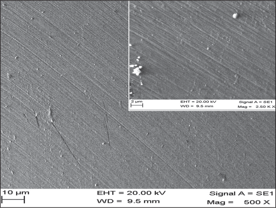
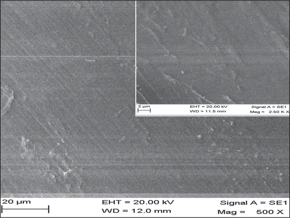
Scanning electron microscope analysis of fluorosed enamel [Figure 1] which served as the control tooth showed pitted, uneven and rough surface with scratches. When samples from Group 1 in which finishing was done with TCBs were evaluated under SEM [Figure 2], the enamel surface was found to have an irregular and rough enamel surface showing horizontal scratches with a consistent pattern with adhesive remnants left on the surface. SEM evaluation of samples from Group 2 [Figure 3] revealed that sof-lex aluminum oxide abrasive discs showed a significant amount of decrease in surface irregularities but fine scratches were seen in every direction. Samples from Group 3 in which polishing was done with Pogo one-step micro polishers revealed a surface with the least amount of scratches with very few remnants on the surface [Figure 4]. When samples which were finished with a combination of TCBs and sof-lex aluminum oxide abrasive discs from Group 4 were subjected to SEM evaluation [Figure 5], it was found that this method was not efficient in removing the scratches on the enamel surface produced by the bur. In Group 5, the samples which were finished with Pogo micro polisher after TCB, the enamel surface had residual scars produced by the bur which could not be efficiently removed by the micro polisher system [Figure 6].
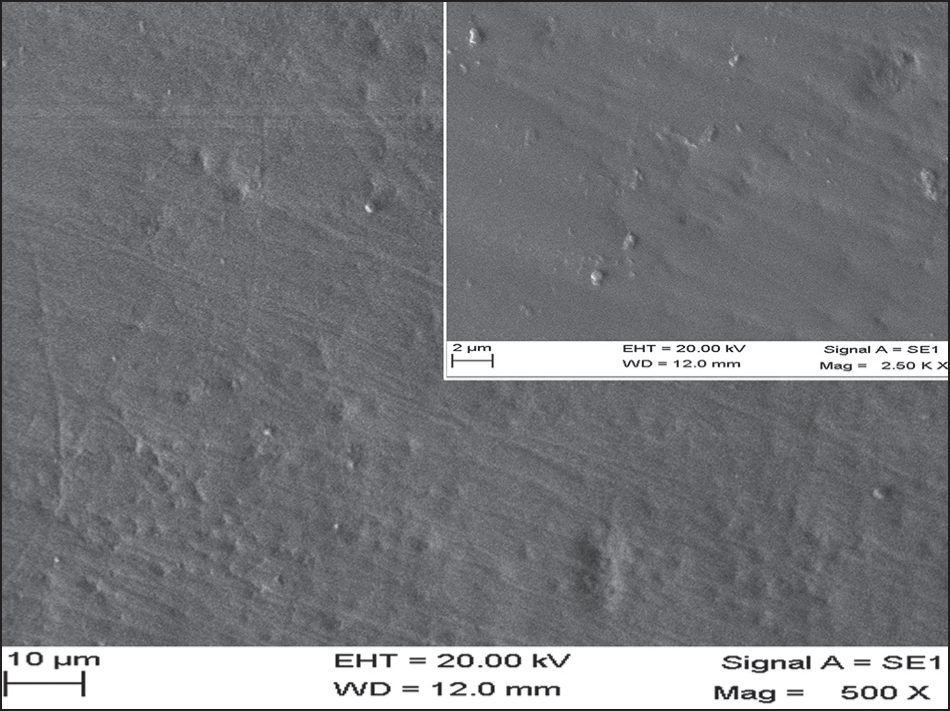
- Scanning electron microscope picture of control tooth at ×500 and ×2500 magnification
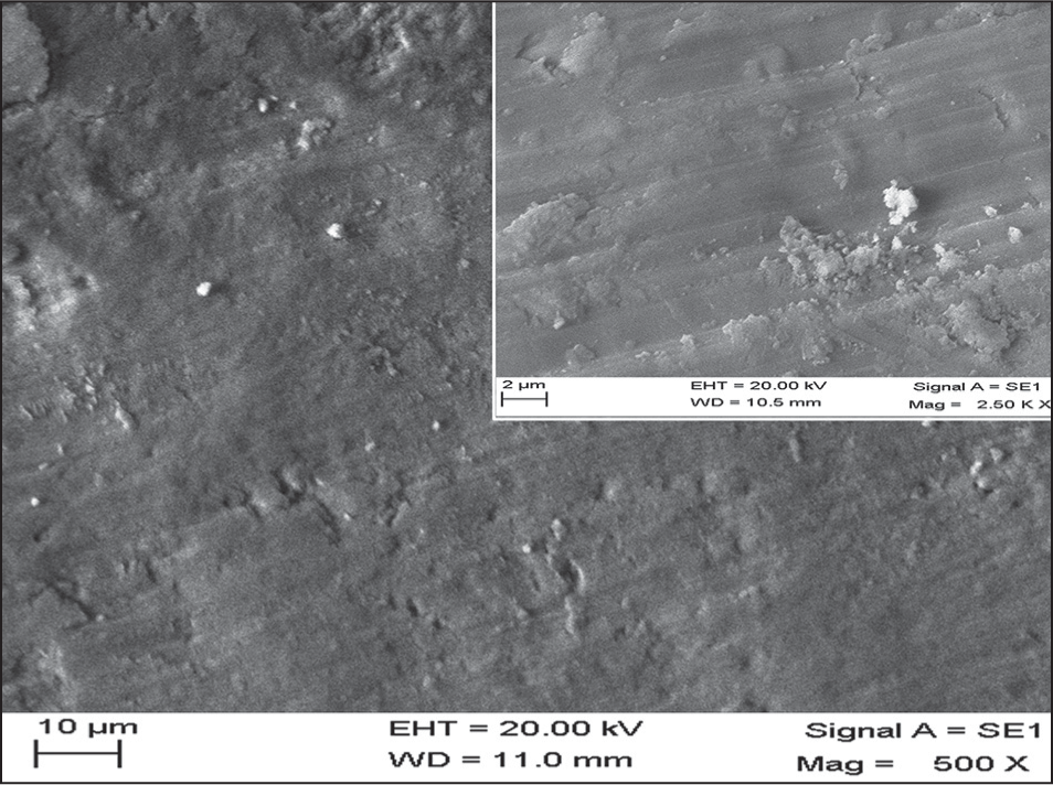
- Scanning electron microscope picture of tooth surface finished with tungsten carbide bur at x500 and x2500 magnification
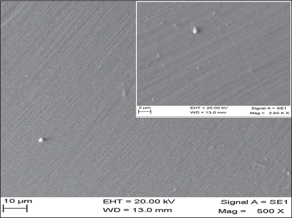
- Scanning electron microscope picture of tooth surface finished with sof-lex discs at x500 and x2500 magnification
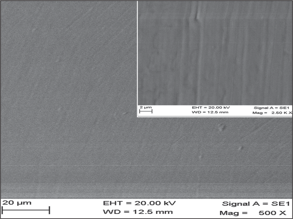
- Scanning electron microscope picture of tooth surface finished with PoGo discs at x500 and x2500 magnification
- Scanning electron microscope picture of tooth surface finished with tungsten carbide bur and sof-lex discs at x500 and x2500 magnification
- Scanning electron microscope picture of tooth surface finished with tungsten carbide bur and PoGo discs at x500 and x2500 magnification
DISCUSSION
Orthodontic treatment has a profound influence on the enamel surface during and after treatment. Subsequent to debonding, the residual adhesive is removed to produce a surface that closely resembles the pretreatment enamel surface. The outermost layer of enamel is highly mineralised and has greater fluoride content; hence this layer serves to protect the surface from decalcification. Irrespective of the technique used for removal of the residual adhesive on the tooth surface, some amount of damage, enamel loss and scarring of the surface is unavoidable.[3]
Metal brackets were preferred to ceramic brackets as they have shown to produce enamel cracks and fractures.[23] The teeth selected for the study were in the mild to moderate score of the modified Deans index. The enamel surfaces of these teeth were hypermineralised due to fluorosis. It was found that all the finishing techniques used in the study produced an acceptable enamel surface to the naked eye; however, on SEM evaluation, each technique was found to have produced varying degrees of roughness and scarring. SEM evaluation of enamel provides an assessment of the enamel surface smoothness and has been used in several studies.[4,8,9,11,15-,16,17] Although SEM can only provide nonquantitative data and subjective information,[24] it is very useful for evaluation of enamel surface topography after various finishing and polishing procedures. In the present study, SEM has been used for evaluation of the enamel surface after different polishing procedures.
After bracket debonding, the mechanical removal of the residual adhesive using rotary instruments leads to enamel loss. TCB’s have been recommended for removal of adhesive in several studies, both in slow speed and at high speed. Hosein et al.[5] in 2004 reported that high speed TCBs produced the most amount of enamel loss in comparison to slow speed burs or debonding pliers. TCB’s are marketed in various shapes, sizes and grits. The 12 fluted bur has been used successfully for adhesive removal by Rouleau et al.[8] In the present study, the 12 fluted TCB at low speed was used with adequate air cooling. SEM pictures (Group 1) of the enamel surface after finishing procedure revealed extreme scarring with a highly rough enamel finish. Retief and Denys,[4] van Waes et al.,[14] Rouleau et al.[8] and Eminkahyagil et al.[15] have also found similar enamel surface after finishing with burs.
Zarrinnia et al.[17] suggested the use of high speed TCB followed by sof-lex discs. These discs are available in various grits namely coarse, medium, fine and ultrafine and are coated with aluminium-oxide particles. Sequential use of these multi-step finishing discs was recommended to produce an enamel surface as smooth as possible. In the present study, sof-lex discs were used subsequent to the initial use of a TCB which removed the bulk of the adhesive (Group 4). Although the time required to complete the finishing procedure was longer than Group 1, SEM evaluation revealed that the enamel surface had lesser surface irregularities. These findings were similar to Zarrinnia et al.[17] However, when sof-lex discs were used alone as in Group 2, the enamel surface had even lesser surface irregularities; but the procedure required a longer period of time to produce that smooth appearance on SEM.
Recently one-step polishers have been introduced which are manufactured with diamond or silicon carbide particles. These have been marketed as the Pogo one-step micro polishing systems. These are designed for a single use without water for the final polishing of composite restoration.[25-27] St-Georges et al.[28] used these Pogo micro polishers to finish composite surfaces in restorations. In the present study, Pogo was used along with TCB in Group 5 and alone in Group 3. When Pogo was used along with TCB, the enamel surface appearance under SEM was better than Groups 1, 2 and 4. The surface was smooth with very fine irregularities. The time taken to produce this smooth surface was similar to Group 4. When Group 3, in which the residual adhesive was removed with only PoGo one-step polishers, was evaluated on SEM, the enamel surface appeared to be the smoothest. This was in concurrence with the findings of Ulusoy[20] and Turkun.[25] However, the one-step polishing system required the longest time to achieve the smoothest enamel finish.
Thus, in the present study, the smoothest enamel surface was produced by the finishing systems in the following order: Pogo one-step polishers when used alone >sof-lex discs > Pogo along with TCB >sof-lex along with TCB >TCB alone. The fastest removal of composite was in the following order: TCB > sof-lex along with TCB> Pogo along with TCB >Pogo alone >sof-lex alone. Thus, from the findings of this study, the use of PoGo alone is recommended to produce a smooth enamel surface which resembles the pretreatment enamel as closely as possible even on fluorosed teeth. All the other finishing systems used in the present study also produced an acceptable enamel finish although a certain amount of residual damage to the enamel is inevitable in all the procedures.
CONCLUSIONS
The following conclusions can be drawn from the study:
All the techniques tested for removal of adhesive on fluorosed teeth produced varying degrees of enamel damage.
The smoothest enamel surface was seen in the following order: PoGo one-step polishers when used alone > sof-lex discs > Pogo along with TCB > sof-lex along with TCB > TCB alone.
TCB took the shortest period of time for resin removal whereas PoGo alone took the longest time.
Thus, for removal of adhesive on fluorosed teeth, the one-step micro polishers provide the best enamel surface which resembles pretreatment enamel as closely as possible at the cost of time. However, if chair side time needs to reduced, then one-step micro polishers may be combined with TCBs.
Source of Support:
Nil.
Conflict of Interest:
None declared.
References
- A simple method of increasing the adhesion of acrylic filling materials to enamel surfaces. J Dent Res. 1955;34:849-53.
- [Google Scholar]
- Orthodontic bracket removal using conventional and ultrasonic debonding techniques, enamel loss, and time requirements. Am J Orthod Dentofacial Orthop. 1993;103:258-66.
- [Google Scholar]
- Finishing of enamel surfaces after debonding of orthodontic attachments. Angle Orthod. 1979;49:1-10.
- [Google Scholar]
- Enamel loss during bonding, debonding, and cleanup with use of a self-etching primer. Am J Orthod Dentofacial Orthop. 2004;126:717-24.
- [Google Scholar]
- Microscopic evaluation of enamel after debonding: Clinical application. Am J Orthod. 1977;71:651-65.
- [Google Scholar]
- Enamel loss due to orthodontic bonding with filled and unfilled resins using various clean-up techniques. Am J Orthod. 1980;77:269-83.
- [Google Scholar]
- Enamel surface evaluations after clinical treatment and removal of orthodontic brackets. Am J Orthod. 1982;81:423-6.
- [Google Scholar]
- Enamel surface characteristics on removal of bonded orthodontic brackets. Am J Orthod. 1978;74:176-87.
- [Google Scholar]
- A new carbide finishing bur for bracket debonding. J Orofac Orthop. 2001;62:296-304.
- [Google Scholar]
- Enamel surface appearance after various debonding techniques. Am J Orthod. 1979;75:121-7.
- [Google Scholar]
- Enamel loss at bond-up, debond and clean-up following the use of a conventional light-cured composite and a resin-modified glass polyalkenoate cement. Eur J Orthod. 2005;27:413-9.
- [Google Scholar]
- Three-dimensional measurement of enamel loss caused by bonding and debonding of orthodontic brackets. Am J Orthod Dentofacial Orthop. 1997;112:666-9.
- [Google Scholar]
- Effect of resin-removal methods on enamel and shear bond strength of rebonded brackets. Angle Orthod. 2006;76:314-21.
- [Google Scholar]
- Enamel loss associated with orthodontic adhesive removal on teeth with white spot lesions: An in vitro study. Am J Orthod Dentofacial Orthop. 2004;125:733-9.
- [Google Scholar]
- The effect of different debonding techniques on the enamel surface: An in vitro qualitative study. Am J Orthod Dentofacial Orthop. 1995;108:284-93.
- [Google Scholar]
- Quantitative and qualitative assessment of enamel surface following five composite removal methods after bracket debonding. Eur J Orthod. 1995;17:121-8.
- [Google Scholar]
- The effect of different polishing systems on surface roughness and gloss of various resin composites. J Esthet Restor Dent. 2007;19:214-24.
- [Google Scholar]
- Comparison of finishing and polishing systems for residual resin removal after debonding. J Appl Oral Sci. 2009;17:209-15.
- [Google Scholar]
- A modified DDE Index for use in epidemiological studies of enamel defects. J Dent Res. 1989;68:445-50.
- [Google Scholar]
- Analysis of three dental fluorosis indexes used in epidemiologic trials. Braz Dent J. 1999;10:29-37.
- [Google Scholar]
- The use of debonding pliers in the removal of ceramic brackets: Force levels and enamel cracks. Am J Orthod Dentofacial Orthop. 1995;108:242-8.
- [Google Scholar]
- Is scanning electron microscopy/energy dispersive X-ray spectrometry (SEM/EDS) quantitative? Scanning. 2013;35:141-68.
- [Google Scholar]
- The effect of one-step polishing system on the surface roughness of three esthetic resin composite materials. Oper Dent. 2004;29:203-11.
- [Google Scholar]
- Thermal changes in the pulp chamber during different adhesive clean-up procedures. Angle Orthod. 2005;75:220-5.
- [Google Scholar]
- Finishing/polishing of composite and compomer restoratives: Effectiveness of one-step systems. Oper Dent. 2004;29:275-9.
- [Google Scholar]
- Surface finish produced on three resin composites by new polishing systems. Oper Dent. 2005;30:593-7.
- [Google Scholar]


