
Translate this page into:
A new skeletal retention system for retaining anterior open bites
This article was originally published by Wolters Kluwer and was migrated to Scientific Scholar after the change of Publisher.
How to cite this article: Albaker B, Rabie B, Wong R. A new skeletal retention system for retaining anterior open bites. APOS Trends Orthod 2013;3:49-53.
Abstract
Objectives
Relapse of anterior open bite after treatment poses a challenge to orthodontists and warrants finding new methods. We aimed to compare the effect of a skeletal retention (SR) system to the conventional retention (CR) commonly used.
Materials and Methods
Twenty patients participated in this study. SR group ten patients (five females and five males) with mean age of 16.2 years, CR group ten patients (five females and five males) with mean age of 17.1 years in pretreatment stage. The SR system is comprised of four self-drilling miniscrews and vacuum retainers with interarch elastics where the CR group is comprised of removable or fixed retainers. Pretreatment (T1), posttreatment (T2), and 1-year follow up (T3) lateral cephalograms were taken and analyzed to compare the stability of both retention modalities.
Results
The overbite in the CR group showed more relapse in the form of significant reduction when compared to the SR group (P< 0.001). The overbite was reduced only by 0.1 mm (±0.3) in the SR group compared to 1.4 mm (±0.9) in the CR group. In the CR group, the upper incisors and first molar showed a more significant relapse compared to the SR group (P< 0.05).
Conclusion
Skeletal retention using miniscrews and vertical elastic is an effective method for retention of anterior open bite cases.
Keywords
Miniscrews
open bite
relapse
retention

[SHOW_RELATED_PUBMED_ARTICLES]
INTRODUCTION
Treatment of anterior open bite is considered a difficult treatment procedure together with the subsequent challenging retention,[1] where relapse has been reported in 25% to 38% of conventionally treated and conventionally retained open bite cases.[2]
It is generally agreed that the remarkable vertical development of the posterior teeth causing a mandibular clockwise rotation, combined with the changes at the incisors during and after orthodontic treatment, are the main reasons for the decrease of the anterior overbite after treatment.[3,4] To enhance the stability after treatment, the cause of the open bite should be eliminated[5] and prolonged retention is advisable and necessary in most cases.[6]
Different approaches have been advocated for retention after treatment of open bite. Day time wear of removable retainers and night time wear of either high pull headgear, or functional appliance with bite blocks (an open bite bionator).[7] Others suggested retainers with occlusal coverage to control molar eruption.[6]
Although many studies evaluated the effect of different treatment procedures (extraction, nonextraction, and surgical) of open bite on the stability and the relapse rate,[1-3,5,8-11] there appear to be no studies that evaluated the stability of open bite cases based on different retention modalities.
Miniscrews offer an excellent anchorage method and have been used to manage open bite successfully in many studies.[12-16] Therefore, they may be used as skeletal retention during the retention period to minimize the relapse of open bite cases.
The aim of this study was to introduce a new retention protocol for open bite by using miniscrews combined with intraoral vertical elastics, and to evaluate and compare this retention regime to that of the conventional retention regime in orthodontically treated open bite patients.
MATERIALS AND METHODS
Subjects
The sample consisted of 20 patients (ten females and ten males) with mean age of 16.6 years in the pretreatment stage.
The patients were selected according to the following criteria.
Pretreatment open bite: Open bite was determined by a negative vertical overlap of at least 1 mm assessed on the pretreatment study models.
Positive overbite at the end of the post-treatment period.
Comprehensive orthodontic treatment.
No surgical treatment.
No cleft and palate patients or syndromic patients
The skeletal retention group (SR) group consisted of ten patients (five females and five males) with mean age of 16.2 years in the pretreatment stage. Six patients had Angle Class I malocclusion, and four had Class II malocclusion. Six patients were treated with four first premolar extractions, and four were treated nonextraction. The mean time was 2.4 years between pretreatment (T1) and post-treatment (T2), and 1 year between post-treatment (T2) and follow up (T3).
The conventional retention group (CR) consisted of ten patients (five females and five males) with mean age of 17.1 years in the pretreatment stage. Seven patients had Angle Class I malocclusion, and three had Class III malocclusion. Five patients were treated with four first premolar extractions, one was treated with first molar extractions, and four were treated nonextraction. The mean time was 3.1 years (range, 2.3-3.8 years) between pretreatment (T1) and post-treatment (T2), and 1 year between post-treatment (T2) and follow up (T3).
Ethical approval for the research was obtained from the local research ethics committee. Procedures were explained to patients in details and all patients signed an informed consent. Pretreatment (T1), post-treatment (T2) and 1-year follow up (T3) lateral cephalograms were taken for all 20 patients under standardized procedures.
RETENTION METHOD
Skeletal retention group
Four self-drilling Orlus* miniscrews [1.6 mm × 1.9 mm × (1.0 + 6.0 mm)] were placed in the attached gingiva of the upper and lower alveolus, 4-5 mm apical to the gingival margin, corresponding to the pretreatment open bite region. After successful insertion of the screws, patients were instructed to use vertical elastic, with a force magnitude of 100-150 g, between upper and lower miniscrews on both sides at night time [Figure 1]. This was in conjunction with vacuum formed retainer worn full time during the first 6 months followed by night time wear for the following 6 months.

- The SR system: Miniscrews, vacuum retainer, and elastics
Conventional retention group
Upper and lower removable or fixed retainers were provided to the patients. Patients were instructed for full time wear during the first 6 months followed by night time wear for the following 6 months.
Cephalometric analysis
The lateral cephalograms were analyzed by one observer to compare the vertical changes between the two groups. Five angular measurements and six linear measurements were used in this analysis [Table 1 and Figure 2].
| Angular cephalometric measurements | |
| ML/NL | The angle between the mandibular line (ML) and the nasal line (NL) |
| ML/NSL | The angle between the mandibular line (ML) and the Nasion-Sella line (NSL) |
| NL/NSL | The angle between the Nasion-Sella line (NSL) and the Nasal line (NL) |
| ILs/NSL | The angle between the long axis of the upper incisor (ILs) and the Nasion-Sella line (NSL) |
| ILi/ML | The angle formed by the long axis of the lower incisor (ILi) and the mandibular line (ML) |
| Linear cephalometric measurements | |
| OB | Overbite, the distance between the perpendiculars from the incisal edges of the upper and lower. incisors to the N-Me line |
| U1-NL | Perpendicular distance between incisal edge of maxillary central incisor and nasal line |
| U6-NL | Perpendicular distance between mesial cusp of maxillary first molar and nasal line |
| L1-ML | Perpendicular distance between incisal edge of mandibular central incisor and mandibular line |
| L6-ML | Perpendicular distance between mesial cusp of mandibular first molar and mandibular line |
| LAFH | Lower anterior facial height: Distance from anterior nasal spine to menton |
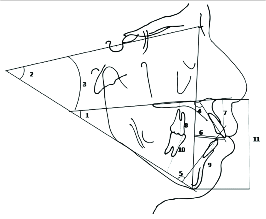
- Angular and linear measurements used in the present study: 1. ML / NL, 2. ML / NSL, 3. NL / NSL, 4. ILs / NSL, 5. ILi / ML, 6. OB, 7. U1-NL, 8. U6-NL,9. L1-ML, 10. L6-ML, 11. LFH
Method error
Ten cephalograms were traced independently on two separate occasions with 2-week interval to determine the method error (ME). The magnitude of the combined ME in locating, superimposing and measuring the changes in different landmarks was calculated by the formula ME = √(Σd2/2n), Where d is the difference between two measurements of pairs of a pair and n is the number of subjects.
The ME for treatment changes did not exceed 0.7 mm for any of the variables investigated.
Statistical analysis
Statistical analysis was carried out using SPSS version 13.0 for Windows (SPSS, Chicago, Illinois, USA) and the descriptive statistics was presented as mean ± SD. Normality test showed that all variable had a normal distribution. Therefore, independent sample t-test was used to compare the pretreatment, post-treatment results between the experimental and control groups. Also it was used to compare the difference between the post treatments and follow up between the two groups.
RESULTS
There was no statistical significant difference at the start of treatment (T1) and post-treatment (T2) between the SR and CR group that made the result comparable [Table 2]. In the SR group, the mean open bite value decreased from –1.9 mm (±0.5) at T1 to 2.0 mm (±0.5) at T2. In the CR group, the mean open bite value decreased from −2.2 mm (±0.9) at T1 to 2.5 mm (±0.8) at T2. In both groups, upper and lower incisors have shown an average retroclination as a result of treatment.
| Variable | Pretreatment | Pvalue | Post-treatment | Pvalue | ||||||
|---|---|---|---|---|---|---|---|---|---|---|
| Experimental group | Control group | Experimental group | Control group | |||||||
| Mean | SD | Mean | SD | Mean | SD | Mean | SD | |||
| Angular(°) | ||||||||||
| ML/NSL | 39.4 | 8.2 | 40.9 | 7.0 | 0.67 | 38.9 | 8.0 | 40.4 | 7.0 | 0.66 |
| NL/NSL | 9.8 | 3.0 | 10.7 | 2.1 | 0.46 | 9.5 | 3.1 | 10.9 | 1.5 | 0.20 |
| ML/NL | 31.5 | 5.3 | 31.2 | 4.6 | 0.89 | 31.1 | 5.7 | 31.4 | 4.6 | 0.90 |
| Uincisor | 120.7 | 6.4 | 120.7 | 8.4 | 1.00 | 111.8 | 6.0 | 110.8 | 7.3 | 0.74 |
| Lincisor | 93.1 | 6.5 | 96.0 | 7.5 | 0.37 | 89.5 | 7.6 | 93.0 | 3.2 | 0.21 |
| Linear (mm) | ||||||||||
| OB | –1.9 | 0.5 | –2.2 | 0.9 | 0.30 | 2.1 | 0.5 | 2.5 | 0.8 | 0.21 |
| U1 | 30.6 | 3.8 | 31.7 | 2.2 | 0.46 | 35.3 | 3.7 | 35.0 | 2.9 | 0.82 |
| U6 | 25.5 | 3.3 | 25.8 | 3.6 | 0.83 | 26.9 | 2.2 | 26.7 | 3.3 | 0.87 |
| L1 | 45.4 | 5.7 | 46.8 | 3.4 | 0.51 | 48.9 | 4.3 | 50.4 | 2.0 | 0.33 |
| L6 | 37.4 | 5.3 | 37.9 | 4.1 | 0.82 | 38.5 | 4.6 | 40.6 | 2.4 | 0.21 |
| LFH | 75.0 | 7.0 | 75.8 | 4.8 | 0.77 | 78.5 | 7.5 | 78.3 | 4.4 | 0.93 |
NSL = Nasion-Sella line; ML = Mandibular line; NL = Nasal line; LFH = Lower facial height; OB = Over bite; SD = Standard deivation
One year follow up
Table 3 shows a comparison of the difference of cephalometric measurements between the follow up (T3) and post treatment (T2) periods in both groups.
| Variable | Experimental group | Control group | Pvalue | ||
|---|---|---|---|---|---|
| Mean | SD | Mean | SD | ||
| Angular(°) | |||||
| ML/NSL | –0.1 | 1.8 | 1.1 | 0.9 | 0.086 |
| NL/NSL | 0.0 | 0.8 | 0.6 | 0.9 | 0.210 |
| ML/NL | 0.1 | 1.9 | 0.9 | 1.4 | 0.298 |
| Uincisor | 0.8 | 1.1 | 3.6 | 2.2 | 0.002* |
| Lincisor | 1.0 | 1.5 | –0.3 | 4.5 | 0.430 |
| Linear (mm) | |||||
| OB | –0.1 | 0.3 | –1.4 | 0.9 | 0.000** |
| U1 | –0.5 | 0.5 | –1.6 | 1.4 | 0.566 |
| U6 | –0.2 | 0.4 | 0.8 | 1.3 | 0.046* |
| L1 | –0.4 | 1.1 | –1.1 | 3.9 | 0.570 |
| L6 | 0.2 | 1.1 | –0.1 | 3.1 | 0.813 |
| LFH | 0.3 | 0.8 | 1.5 | 2.4 | 0.145 |
The overbite in the CR group showed a more significant reduction, that is, more relapse when compared to the SR group (P< 0.001). The overbite was reduced only by 0.1 mm (±0.3) in the SR group compared to 1.4 mm (±0.9) in the CR group [Table 3].
The upper dental measurements were significantly different between the two groups. In the CR group, the upper incisors and first molars showed a more significant relapse compared to the SR group (P< 0.05).
DISCUSSION
In this study, a new protocol for enhancing vertical stability of orthodontically treated open bite patients by skeletal retention through miniscrews was presented and compared with the conventional retention method. The lack of significant difference between pretreatment (T1) and post-treatment (T2) measurements of the two groups enabled the assessment of the results of the retention phase based on the retention modality.
The SR system is comprised of four miniscrews in each arch placed where the open bite existed before treatment. For example, if the open bite spanned between both upper canines, the mini screws would be placed distal to upper canines. Interarch elastics are then prescribed to the patient to wear at night. If the open bite included both right and left upper laterals, then the miniscrews would be placed mesial to upper canines. A vacuum retainer is also fabricated to guard against the unwanted side effects of the interarch elastics as they rest on the teeth.
During the follow-up period, the SR group achieved stability as demonstrated by the reduction of the overbite by only 0.1 mm [Figure 3]. The CR group demonstrated a relapse of 1.4 mm in the overbite in the CR group. Results of the CR are in accordance with the study of Kucukkeles et al. who found a reduction of 1.25 mm of the overbite.[4]

- (a) Post-treatment photo prior to starting the SR. (b) Same patient after 1-year retention with SR
Changes in the maxillary teeth seemed to have played an important role in the overbite changes as the upper incisors showed a significant change due to combination of proclination and intrusion. The upper molars showed a significant degree of extrusion in the CR group compared to the SR group in the follow-up period. Therefore, the SR system, keeping both arches in contact as a result of the use of interarch elastics, could provide enough retention to guard against the eruption of the molars. Furthermore, the use of the vacuum retainers together with the interarch elastics guards against proclination of the incisors and thus provides more stable retention outcome.
Although the increase in the ML/NSL angle, during the follow up period, in the CR group was not statistically significant compared to the SR group, yet the increase of 1 degree in the CR group demonstrated clinical significance manifested in the decrease of overbite by 1.4 mm [Table 3]. The 1 degree of increase in the ML/ NSL indicates a clockwise mandibular rotation after retention, which in turn could contribute to anterior open bite relapse.[2,3]
An obvious advantage of the skeletal retention method in this study is that the timing of treatment of overbite may not be as critical as in cases managed with other means of retention[8,17] since any unfavorable growth direction of the mandible can be well controlled. However, this method does not eliminate the need of continued retention until growth is completed.
So far we have successfully treated a few relapsed cases with the current protocol (unpublished data) which is yet another potential usage for this method to treat relapsed open bite cases in growing patients by redirecting the growth of the mandible to a more favorable pattern. However, further studies are needed to evaluate the treatment response.
The weakness of the current study is the relatively small sample size and the evaluation of only 1 year of retention. Studies with a bigger sample size and longer retention period are being carried out.
CONCLUSION
Skeletal retention using miniscrews and vertical elastic is an effective method for retention of anterior open bite cases.
Source of Support:
Nil.
Conflict of Interest:
None declared.
References
- Long-term stability of anterior open-bitetherapy: A review. Semin Orthod. 2002;8:162-72.
- [Google Scholar]
- Anterior open-bite malocclusion: A longitudinal 10-year postretention evaluation of orthodontically treated patients. Am J Orthod. 1985;87:175-86.
- [Google Scholar]
- Stability of anterior openbite correction with multiloop edgewise archwire therapy: A cephalometric follow-up study. Am J Orthod Dentofacial Orthop. 2000;118:43-54.
- [Google Scholar]
- Cephalometric evaluation of open bite treatment with NiTi arch wires and anterior elastics. Am J Orthod Dentofacial Orthop. 1999;116:555-62.
- [Google Scholar]
- Nonsurgical management of the anterior open bite: A review of the options. Semin Orthod. 1999;5:275-83.
- [Google Scholar]
- Retention. In: Proffit WR, ed. Contemporary Orthodontics (4th ed). St. Louis: Mosby; 2007. p. :622.
- [Google Scholar]
- Anterior open bite malocclusion: A follow-up study of orthodontic treatment effects. Eur J Orthod. 1993;15:273-80.
- [Google Scholar]
- Stability of anterior open bite nonextraction treatment in the permanent dentition. Am J Orthod Dentofacial Orthop. 2003;124:265-76. quiz 340
- [Google Scholar]
- Long-term stability of anterior open bite extraction treatment in the permanent dentition. Am J Orthod Dentofacial Orthop. 2004;125:78-87.
- [Google Scholar]
- Stability of anterior open-bite extraction and nonextraction treatment in the permanent dentition. Am J Orthod Dentofacial Orthop. 2006;129:768-74.
- [Google Scholar]
- Severe anterior open-bite case treated using titanium screw anchorage. Angle Orthod. 2004;74:558-67.
- [Google Scholar]
- Treatment and posttreatment dentoalveolar changes following intrusion of mandibular molars with application of a skeletal anchorage system (SAS) for open bite correction. Int J Adult Orthodon Orthognath Surg. 2002;17:243-53.
- [Google Scholar]
- Skeletal anchorage system for open-bite correction. Am J Orthod Dentofacial Orthop. 1999;115:166-74.
- [Google Scholar]
- Closing anterior open bites by intruding molars with titanium miniplate anchorage. Am J Orthod Dentofacial Orthop. 2002;122:593-600.
- [Google Scholar]
- The use of skeletal anchorage in open bite treatment: A cephalometric evaluation. Angle Orthod. 2004;74:381-90.
- [Google Scholar]


